What is Retinal Detachment and Various Types of Surgeries?
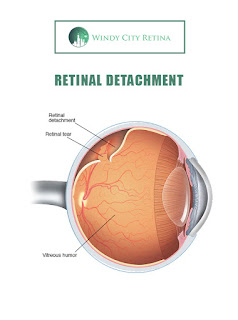
Retinal detachment is a medical state of the eye when the thin layer of tissue (retina) drags itself from the back of the organ. In medical terms, it is called retinal detachment. Retinal cells are really important for providing an adequate supply of oxygen and nourishment to blood vessels. A serious loss of vision can occur in the absence of any treatment.
There are some people who are at more risk of developing it than others. People who are over the age of forty or people with a history of the same illness in the family are at more risk of developing it than others. People who suffered from eye injuries or with nearsighted vision can also be at the risk, they should get their sight of eyes monitored with a doctor at regular intervals.
There are few symptoms you can experience as warning signs before developing the retinal detachment
1. Flashes of light.
2. Experience of increase in the floaters in the field of
vision.
3. Blurred vision and curtain-like shadow in the field of
vision.
4. Vision reducing gradually.
Retinal detachment is a painless medical condition. You should be aware of these symptoms and take proper measures and treatments before developing this eye condition.
Types of retinal detachment
1. Tractional- In this type of retinal detachment the scar tissue
pulls from the retina and causing the retina to drag away from the organ.
Mainly happen to people with uncontrolled diabetes.
2. Rhegmatogenous- It results from a hole in the retina. The hole allowed the fluid to accumulate underneath the retina and dragging the retina away from the back of the eye. As you age the vitreous gel becomes more liquid and pulls away the retina.
3. Exudative- In Exudative detachment, the fluid gets accumulated underneath the retina but without any tearing or hole. This condition can happen because of an eye injury, inflammatory disorder or any age-related macular degeneration.
Types of treatments of Retinal detachments-
1. Scleral buckle- In this treatment, the doctor heals the tearing or detachment of the retina by sewing the silicone buckle with sclera the white of the eye. It is done with an invisible band to push the retina back to its original location to heal. This procedure can take place under local anaesthetic and has over a 90% success rate.
2. Vitrectomy- In this surgery, the vitreous gel is replaced by a gas bubble or oil by the doctor for healing the detachment. You need to hold your head in one side for proper healing.
3. Pneumatic retinopexy- In this procedure, a bubble of gel is injected into the eye area for healing the tear by pressing the upper part of the retina. It requires your head to be in specific spots so that bubble can heal the retinal detachment.
At last, we can say that we should be aware of symptoms
and take proper actions before developing retinal detachment and preventing
further loss of vision. That is the reason why acting as soon as possible is
recommended.


